



Sleep is essential for our daily lives and consists of two main stages: rapid eye movement (REM) sleep and non-REM sleep. However, it is still not fully understood which neurons control each stage or how these processes are regulated in the brain. To address this question, Assistant Professor Kashiwagi is studying the neurons that regulate sleep using mouse models.

Exploring Brainstem Neural Circuits
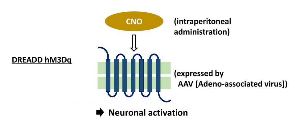
Since my third year as an undergraduate, I have been studying REM sleep in the laboratory of Dr. Yu Hayashi at the International Institute for Integrative Sleep Medicine, University of Tsukuba. In the brain, many different types of neurons work together to control sleep and wakefulness. Among these, REM sleep remains one of the least understood stages. Today, several types of neurons that promote REM sleep have been identified. However, when this research began, such neurons had not yet been clearly identified. To overcome this challenge, we used genetically modified mice that make it possible to identify neurons with specific functions. The brainstem contains many different types of neurons and plays an important role in sleep regulation. However, the function of each group of neurons is still not fully understood. To study this in more detail, we used genetic techniques that allow us to label and control specific neurons. This made it possible to analyze the roles of individual neurons more precisely. To examine how these neurons affect sleep, we used a technique called DREADD (Designer Receptors Exclusively Activated by Designer Drugs). By introducing special receptors into specific neurons using a viral vector and administering a corresponding drug, we were able to either activate or suppress those neurons (Figure 1).
Figure 1. Manipulation of neuronal activity using the DREADD method Artificial receptor (modified from human acetylcholine receptor) Neuronal activation is induced by administration of CNO (clozapine-N-oxide)
(Armbruster, et al., PNAS, 2007)(Created by Mitsuaki Kashiwagi).
To understand how specific neurons influence sleep, we recorded brain and muscle activity in mice using electroencephalography (EEG) and electromyography (EMG). Through these experiments, we identified a group of neurons in the brainstem that suppress REM sleep, known as Nts neurons (Figure 2). These neurons produce a molecule called neurotensin, and we found that neurotensin itself can also suppress REM sleep. In addition, neurotensin-producing neurons located in different parts of the brainstem appear to work together to maintain the balance between REM and non-REM sleep. Interestingly, this neural circuit overlaps with regions involved in sensing body movement. This finding may help explain why gentle motion can make us sleepy—for example, why rocking helps babies fall asleep or why people often feel drowsy when riding on a train.
Figure 2. Left: Image of activated Nts neurons. Nts neurons are visualized in yellow using a fluorescent protein.
Kashiwagi, M. et al. Widely Distributed Neurotensinergic Neurons in the Brainstem Regulate NREM Sleep in Mice. Current Biology, 2020; 30, 1002-1010.e4. Copyright: 2020 Elsevier Ltd. https://doi.org/10.1016/j.cub.2020.01.047
Right: Comparison of sleep patterns between the control condition (normal sleep) and after activation of Nts neurons. Gray indicates wakefulness, light blue indicates non-REM sleep, and purple indicates REM sleep. When Nts neurons are activated, REM sleep (shown in purple) is suppressed compared with the control (normal) condition. (Created by Mitsuaki Kashiwagi).
Identifying Neurons that Promote REM Sleep
Next, we investigated neuronal populations that promote REM sleep. By carefully reviewing previous studies and applying new analytical approaches, we were able to identify—for the first time—neurons in the pons that induce REM sleep (Figure 3). In the brain, neurons communicate with one another in a relay-like manner to control complex functions. We also discovered that neurons capable of inducing REM sleep are present in the medulla, a region located next to the pons. These findings suggest that neurons distributed across the pons and medulla work together to regulate REM sleep.
Figure 3. Discovery of neurons in the pons that induce REM sleep. Representative sleep patterns are shown. Gray indicates wakefulness, light blue indicates non-REM sleep, and orange indicates REM sleep. Activation of neurons in the pons increases REM sleep, as indicated by the increased orange segments.
Reproduced and modified from Kashiwagi, M. et al. A pontine-medullary loop crucial for REM sleep and its deficit in Parkinson’s disease. Cell, 187 (22), 6272 (2024). Copyright 2024 Kashiwagi, M. et al.
Exploring the Mechanisms of REM Sleep as a Global Research Leader
REM sleep is thought to be closely related to psychiatric and neurodegenerative disorders. However, compared to non-REM sleep, its underlying mechanisms are still not well understood. A study demonstrating a neural circuit in which neurons in the pons and medulla cooperate to promote REM sleep was published in the international journal Cell and has attracted considerable attention. Building on these findings, future research will focus on how these neurons are activated and how they interact with other circuits that regulate sleep and wakefulness. Ultimately, this work aims to deepen our understanding of how REM sleep is controlled in the brain and how its disruption may contribute to psychiatric and neurodegenerative diseases.
(Date of interview: February 2, 2026)